best time for 3d ultrasound with anterior placenta
Plan something enjoyable with friends or your partner. As the weeks go on and the baby gets bigger the movements will be more recognizable.
Anyone Had 4d Scan With Anterior Placenta
Academic Radiology provides authors with quick turnarounds throughout the publication process.

. The pain may be described as sharp dull or crampy. Ectopic pregnancy is a complication of pregnancy in which the embryo attaches outside the uterus. 226 Issue 2 p245e1.
Talk to your doctor about any new symptoms including dizziness. If the cord inserts to the margin of the placenta then it should be reported as a marginal insertion and it is helpful to specify the side of. Breast MRI is better than clinical assessment mammogram and ultrasound in correlation with pathology.
Echoes from the various boundaries are given against total time ie. CT is still the choice as the first imaging modality in acute stroke institutional protocols not only because the availability and the easy and fast access to a CT scanner but also due the better sensitivity for intracerebral hemorrhage ICH diagnosis 1. Get ready for your mid-pregnancy ultrasound.
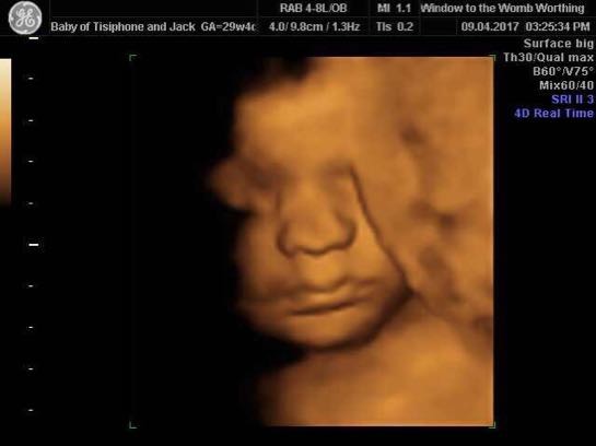
We recommend the best time for 4D is between 26-30 weeks. By 30 weeks babies spend most of their time sleeping and while. Pain may also spread to the shoulder if bleeding into the abdomen has.
Surgical coaching in Obstetrics and Gynecology. Plus-size pregnant women or women whose placenta is on the front wall of the uterus called an anterior placenta may take longer to feel the kicks. Some pregnant people dont feel the baby move until 20 weeks or even later.
It offers diagnostic and imaging services such as ultrasound x-ray mri ct scan and blood tests etc. Other factors that may affect the quality of the 3D4D images are BMI of mummy this is because ultrasound uses sound waves that have to travel through the tissue the higher the amount of tissue the further they have to travel therefore the less clearer the image will be. 47 This is easy with an anterior placenta but can be difficult to locate if the fetus is lying on a posterior cord insertion.
General Sonography Lab II. American Journal of Obstetrics Gynecology Vol. Columns of the fornix arrow.
It is advisable to scan through the placenta to attempt to find where the cord inserts into the placental mass. Continue reading Video of a D E Abortion GraphicIts common for women to push out. It can take a little longer to feel movements for all expecting parents especially for first-time pregnancies or those with an anterior placenta.
Especially if it is a 3D ultrasound. FIGURE 18-6 A. See the months best real-time 3D models from free characters to entire miniature animated worlds.
If you have an anterior placenta 26-28 weeks to ensure theres plenty of room for baby to move. Pretreatment MRI should be performed and compared with MRI done after neoadjuvant chemotherapy this is particularly useful to monitor response to neoadjuvant chemotherapy allowing to identify non-responders early and to delineate the residual tumor. 2016 segmented nineteen targets in the human torso and Moeskops et al.
If they have an anterior placenta where the placenta is at the front it will also mean they may need to wait a bit longer. Fast Decision and Publication Time. This was an original video but it was taken off youtube by people who want to censor the truth.
Intrauterine devices IUDs are a commonly used form of contraception worldwide. DMS207 75 hours This sonography course is designed to offer the student of sonography an opportunity to practice techniques learned in. An ultrasound photo is a 2D image of a 3D place.
Your Week 18 Checklist. The important thing is to take time to do what makes you happy. Moolchand is the best hospital chain in Delhi Agra with specialist doctors - intensivists nephrologists gastroenterologists general surgeons cardiologists neurologists orthopaedicians paediatricians gynaecologists emergency medicine specialists and more.
Theres a lot to prepare for before the baby. We return initial decisions about a month after receiving a submission on average. Excessive Opioid Prescribing After Major Urologic Procedures.
In diagnosis it is used to create an image of internal body structures such as tendons muscles joints blood vessels and internal organs to measure some characteristics eg. Free Ambulance Services Reports Panel - 011 4744-4444 011 4733-3333 Online Reports. 2016b trained a single fCNN to.
FCNNs have also been extended to 3D and have been applied to multiple targets at once. Signs and symptoms classically include abdominal pain and vaginal bleeding but fewer than 50 percent of affected women have both of these symptoms. For mutiple pregnancies we recommend visiting us from 22 - 26 weeks.
Its the inside of your uterus. The velocity of the ultrasound waves in the eye is approximately 1550 ms it is 1641 ms in the lens and 1532 ms in the humours. The length is then equal to.
Advice for Partners. Single-center experience with a multidisciplinary team. If your placenta is anterior at the front we recommend you try and come before 29 weeks to avoid.
Indias 1st JCI and NABH accredited Hospital. Lower uterine segment contractions should be considered whenever the cervical length measures more than 50 mm the cervical canal assumes an S shape and the lower uterine segments either anteriorly or. The septum pellucidum is part of the limbic system and has connections to the hypothalamus hippocampus and amydala 45The CSP reaches its adult configuration by 17 weeks gestation 56The CSP can be documented in virtually 100 of cases between 18 and 37 weeks gestation.
In the above diagram the total time between the cornea and the retina is 32 μs. If accepted articles are posted online in fully citable form in about 6 weeks and published in a print issue about 4 months after acceptance. Medical ultrasound includes diagnostic techniques mainly imaging techniques using ultrasound as well as therapeutic applications of ultrasound.
Email protected email protected. An evidence-based strategy to elevate surgical. MRI protocol for stroke assessment is a group of MRI sequences put together to best approach brain ischemia.
Distances and velocities or to generate an informative. Urologic morbidity associated with placenta accreta spectrum surgeries. The time interval from the cornea to the boundary and back to the cornea.
If a mom has an appointment coming up and she is anxious to see her little one hopping around there is a good trick she can try. Adapting Urology Residency Training in the COVID-19 Era. The technicians that read your scan for the Ramzi Method like The Gender Experts look for the following.
In a transverse ultrasound a technician can easily tell which side of your body is which based on where the beam enters the body and can annotate your printed ultrasound photos accordingly. Other people can feel your babys movements by placing their hand on your belly but not until a few weeks after youve felt movement from the inside. Streetwear Trendy Stylish Fashion ClothingFootwear More.
2016 used 3D fCNNs to generate vertebral body likelihood maps which drove deformable models for vertebral body segmentation in MR images Zhou et al. Different sites of IUD translocation vary in terms of their clinical. The role of ultrasound in the assessment of the placenta umbilical cord amniotic fluid and membranes and fetal organ systems is included.
General Sonography Lab II. It wont be long before you and loved ones can feel and. The diagram on the left and the ultrasound image on the right show a cervix with significant lower uterine segment contractions asterisks.
Teleurology in the Time of Covid-19 Pandemic. However migration of the IUD from its normal position in the uterine fundus is a frequently encountered complication varying from uterine expulsion to displacement into the endometrial canal to uterine perforation.

3d 4d Ultrasound W Anterior Placenta January 2019 Babies Forums What To Expect

How To Get The Best Ultrasound Photos Mother Nurture Ultrasound

Anyone With Anterior Placenta Have 3d 4d Ultrasound Babycenter

3 4d Ultrasounds Not Great Because Of Anterior Placenta December 2017 Babies Forums What To Expect

3d 4d Ultrasound At 32 Weeks Anterior Placenta March 2019 Babies Forums What To Expect

3d Ultrasounds And Anterior Placenta January 2019 Babies Forums What To Expect

3d Ultrasound And Anterior Placenta August 2018 Babies Forums What To Expect

32 Weeks Anterior Placenta 3d Ultrasound Babycenter

Ultrasound Faqs Baby S First Images Ultrasound